Loose Body Removal

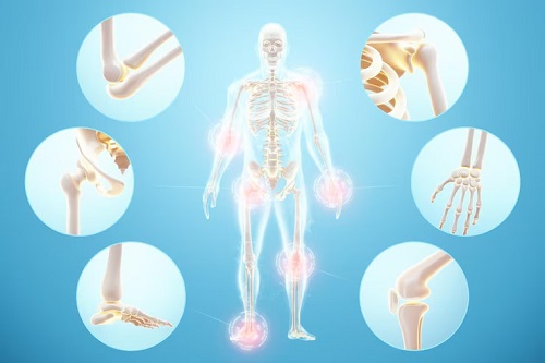
Loose bodies refer to small fragments of bone or cartilage that become detached within the body, leading to potential joint interference or obstruction.
The optimal approach to treating and surgically removing these loose bodies depends on several crucial factors: the individual’s age, overall health history, the size and location of the loose body, as well as any prior medical interventions or medications. Common triggers for loose bodies include sports-related injuries or occupational incidents.
Additionally, degenerative conditions like osteoarthritis often necessitate loose body removal, particularly in individuals who subject their joints to repetitive stress, such as athletes or those engaged in heavy lifting tasks, especially overhead movements.
Types of loose body removal
Nonsurgical Options: In milder cases, physiotherapy and anti-inflammatory medications can offer relief and maintain joint flexibility and mobility. While these methods may alleviate symptoms, regular monitoring and follow-up with a healthcare provider are crucial to prevent deterioration of the condition.
Surgical Intervention: Typically, the removal of loose bodies requires a surgical approach. The goal of this surgery is to address the mobility restriction caused by free-floating cartilage or bone fragments resulting from injuries.
The two primary surgical techniques employed are:
Arthroscopy: This minimally invasive procedure is favored by many physicians. It involves making a small incision through which a camera is inserted, providing visualization of the affected area. The loose bodies are then removed using a suction cup introduced through another small incision.
Arthrotomy: In rare cases where arthroscopy is not feasible, particularly with larger loose bodies, a more extensive surgical procedure called arthrotomy may be necessary. This technique involves a larger incision to improve visibility inside the joint, facilitating easier removal of the loose bodies. Both approaches aim to restore joint function and alleviate symptoms associated with loose bodies, with the choice of technique dependent on factors such as the size and location of the loose bodies and the patient’s overall health.
Diagnosis
- X-ray (radiograph): Uses radiation to create a black-and-white image of the affected joint, allowing for clear visualization of bone and cartilage fragments.
- Computed tomography scan (CT or CAT scan): Combines X-ray and computer technology to produce a high-definition image, offering detailed views of bone structures.
- Magnetic resonance imaging (MRI): Utilizes electromagnetic radio waves to generate detailed images without radiation, particularly useful for detecting non-bony particles.
- Arthrography: Involves injecting dye into the joint area and taking an X-ray to highlight soft tissue and cartilage, offering enhanced detail, especially in smaller joints like the wrist.
